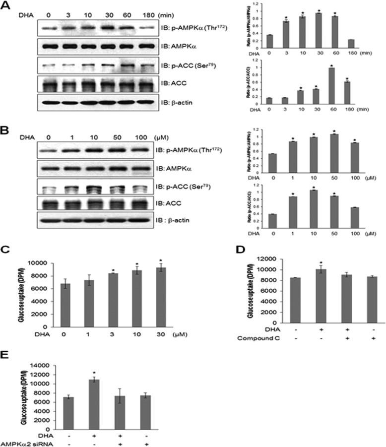
FIGURE 1.
DHA stimulates glucose uptake through AMPK. A, cells were stimulated with DHA for the indicated times and were analyzed by Western blotting with antibodies against phospho-AMPK and phospho-ACC. AMPK and ACC served as controls. The results are representative of four independent experiments. *, p < 0.05 versus basal conditions. B, cells were stimulated with various doses of DHA, and cell lysates were analyzed by Western blotting with antibodies against phospho-AMPK and phospho-ACC. AMPK and ACC served as controls. The results are representative of four independent experiments. *, p < 0.05 versus basal conditions. C, L6 myoblasts were differentiated and stimulated with various doses of DHA for 1 h, and 2-DG uptake was assayed. *, p < 0.05 compared with control. D, L6 myoblasts were differentiated and stimulated using 50 μm DHA for 1 h in the presence of compound C, and 2-DG uptake was assayed. *, p < 0.05 compared with DHA-treated cells. E, L6 myoblasts were transiently transfected with AMPKα2 siRNA for 2 days and stimulated using 50 μm DHA for 1 h, and 2-DG uptake was assayed. *, p < 0.05 compared with control. IB, immunoblot.

