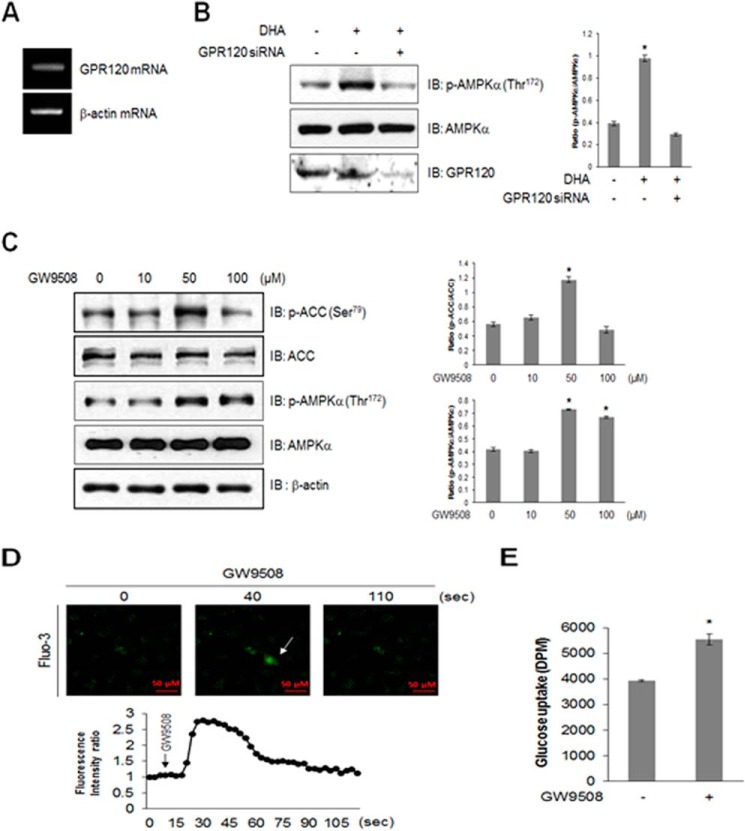
FIGURE 3.
GPR120 mediates DHA-induced AMPK phosphorylation. A, total mRNA was extracted from C2C12 cells, and RT-PCR was performed using GPR120-specific primers. PCR products were visualized under ultraviolet light. β-Actin was used as a positive control. B, C2C12 cells were transiently transfected with GPR120 siRNA for 48 h and stimulated with 50 μm DHA. Cell lysates were analyzed by Western blotting with antibodies against phospho-AMPKα, with AMPKα as a control. The results are representative of four independent experiments. *, p < 0.05 versus basal conditions. C, the cells were stimulated with GW9508 for indicated doses for 1 h, and cell lysates were analyzed using Western blotting with antibodies against phospho-AMPKα and phospho-ACC, with AMPKα, ACC, and β-actin as controls. The results are representative of four independent experiments. *, p < 0.05 versus basal conditions. D, C2C12 cells were pretreated with fluo-3 AM for 30 min and then with 100 μm GW9508. Green fluorescence was detected using confocal microscopy. E, L6 myoblasts were differentiated and stimulated with 50 μm GW9508 for 1 h. Uptake of 2-DG was assayed. *, p < 0.05 compared with control. IB, immunoblot.

