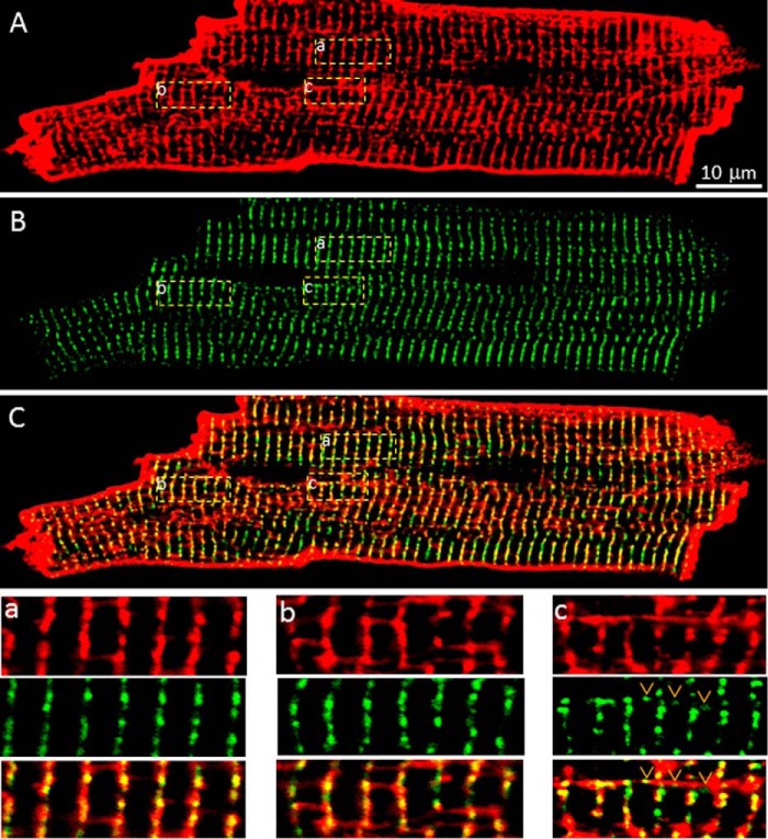
FIGURE 5.
Relative distribution of GFP-RyR2 clusters and the tubular system. A, the tubular membrane system in live GFP-RyR2 ventricular myocytes (n = 27) was labeled with di-8-ANEPPS and visualized with confocal fluorescence imaging. B, GFP-RyR2 cluster fluorescence signals of the same cell. C, merged image of the di-8-ANEPPS and GFP-RyR2 signals. The panels at the bottom show co-localization of GFP-RyR2 clusters with transverse tubules (panel a), no co-localization of GFP-RyR2 clusters with longitudinal tubules (panel b), and co-localization of a few GFP-RyR2 clusters with longitudinal tubules located near the perinuclear region (panel c).

