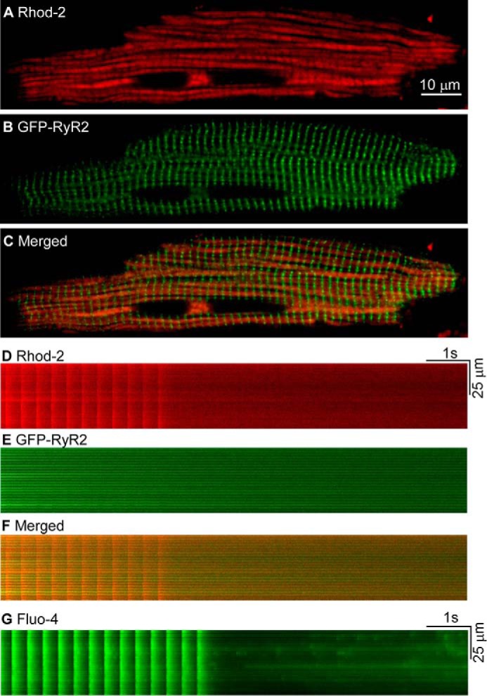
FIGURE 9.

Influence of depolarization-induced Ca2+ release and Ca2+ sparks on mitochondrial Ca2+ level in GFP-RyR2 ventricular myocytes. A, mitochondria in live GFP-RyR2 ventricular myocytes (n = 16) were loaded with Rhod-2 AM and visualized with confocal fluorescence imaging. B, GFP-RyR2 cluster fluorescence signals of the same cell as in A. C, merged image of the Rhod-2 labeling and GFP-RyR2 signals. D, fluorescence signals of Rhod-2 loaded GFP-RyR2 ventricular myocytes (n = 28) during and after termination of pacing at 3 Hz in the presence of 3 mm extracellular Ca2+. E, GFP-RyR2 cluster fluorescence signals of the same cell as in D. F, merged image of the Rhod-2 and GFP-RyR2 signals. G, cytosolic Ca2+ transients and Ca2+ sparks revealed by Fluo-4 in GFP-RyR2 ventricular myocytes (n = 16) during and after termination of pacing at 3 Hz in the presence of 3 mm extracellular Ca2+.
