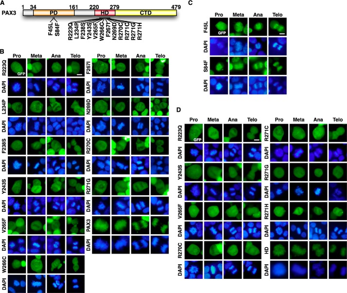
FIGURE 2.
PAX3 HD mutants found in WS lost the ability to load on mitotic chromosomes. A, schematic diagram of PAX3 mutations found in WS patients. B, PAX3 WS mutants in HD do not load on mitotic chromosomes. The expression vectors for GFP-PAX3 WS mutants in HD were transfected in HEK 293 cells. Localization to the mitotic chromosome was determined by fluorescence microscopy. GFP-PAX3 served as control. Meta, metaphase; Ana, anaphase; Telo, telophase. C, PAX3 WS mutants in PD retained the ability to load on mitotic chromosomes. The expression vectors for GFP-PAX3 WS mutants in PD were transfected in HEK 293 cells. Localization to the mitotic chromosome was determined by fluorescence microscopy. D, HD of PAX3 with WS mutations lost mitotic chromosome localization. GFP-tagged HDs harboring mutations found in WS patients were expressed and analyzed for chromosomal localization as described in panels B and C. GFP-HD served as the positive control.

