
FIGURE 10.
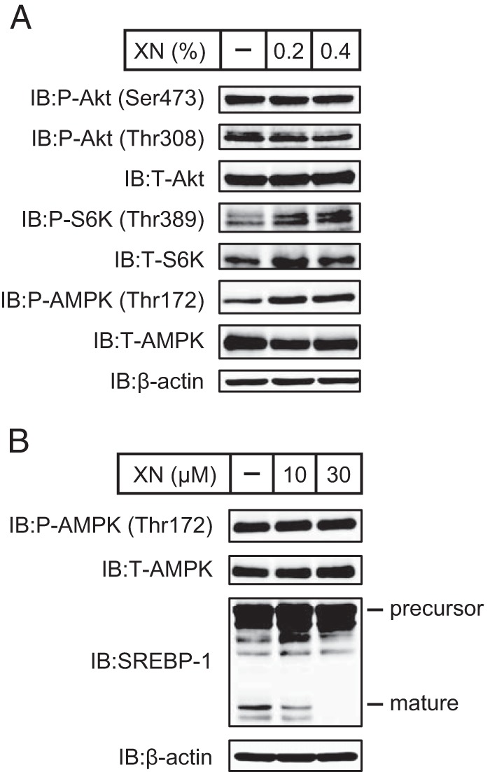
Phosphorylation states of proteins involved in insulin signaling in livers of mice fed a HFD supplemented with XN and XN do not affect AMPK phosphorylation in cells. A, immunoblot (IB) analysis of liver extracts with anti-phospho-Akt (Ser-473), anti-phospho-Akt (Thr-308), anti-Akt, anti-phospho-S6K (Thr-389), anti-S6K, anti-phospho-AMPK (Thr-172), anti-AMPK, or anti-β-actin antibodies. The mice used here were the same as those used in Fig. 9. B, CHO-7 cells were depleted of sterols by incubating in medium F for 16 h. The cells were then switched to medium F in the presence of vehicle, 10 μm XN, or 30 μm XN. After incubation for 3 h, whole cell extracts were subjected to immunoblotting with anti-phospho-AMPK (Thr-172), anti-AMPK, anti-SREBP-1 (2A4), or anti-β-actin antibodies. The same results were obtained in more than three separate experiments.