
FIGURE 5.
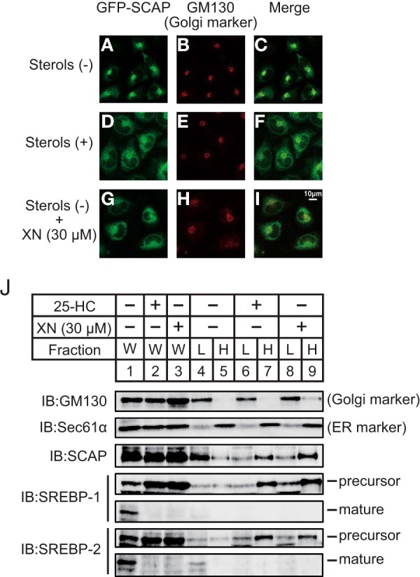
XN blocks the ER-to-Golgi translocation of the SCAP/SREBP complex. A–I, CHO/pGFP-SCAP cells were depleted of sterols by incubating in medium F for 3 h. The cells were then switched to medium F in the absence or presence of sterols (10 μg/ml cholesterol plus 1 μg/ml 25-HC) or 30 μm XN. After incubation for 3 h, the cells were fixed and then stained with primary antibody against GM130 followed by Cy3-conjugated secondary antibody. The cells were then imaged for GFP-SCAP (A, D, and G) and GM130 (B, E, and H). C, F, and I are merged images of GFP-SCAP and GM130. J, CHO-7 cells were depleted of sterols by incubating in medium F for 16 h. The cells were then switched to medium F in the presence of vehicle, 1 μg/ml 25-HC, or 30 μm XN. After incubation for 3 h, the cells were fractionated into a light fraction, containing the Golgi, and a heavy fraction, containing the ER, by sucrose density-gradient centrifugation, as described under “Experimental Procedures.” Equal volumes of each fraction were subjected to immunoblotting (IB) with anti-GM130, anti-Sec61α, anti-SCAP, anti-SREBP-1 (2A4), or anti-SREBP-2 antibodies. The same results were obtained in more than three separate experiments. W, whole cell lysates; L, light fraction; H, heavy fraction.