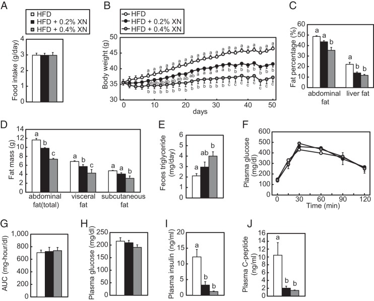
FIGURE 9.
XN inhibits the development of obesity and fatty liver in HFD-fed mice. A–D and F–J, 6-week-old C57BL/6J male mice were fed a HFD for 10 weeks and subsequently fed a HFD, 0.2% XN, or 0.4% XN-supplemented HFD for 50 days (n = 10–11). The mice were given free access to water and their diet. CT scans and OGTT were performed at 35 and 44 days after the initiation of XN supplementation, respectively. The mice were sacrificed and subjected to various analyses as described below. A, total daily average mouse food intake for each treatment group during the 50-day experiment. B, average mouse body weight for each treatment group during the 50-day experiment. C, fat percentages in the abdomen and liver after 35 days of XN feeding. Mice were scanned using an experimental animal x-ray CT system (LaTheta LCT-200). D, abdominal fat mass after 35 days of XN feeding. Mice were analyzed as in C. Whole abdominal fat, visceral fat, and subcutaneous fat masses were determined using CT analysis. E, feces triglyceride levels for each treatment group. Six-week-old C57BL/6J male mice (n = 5) were fed a HFD for 1 week and subsequently fed a HFD, 0.2% XN, or 0.4% XN-supplemented HFD for 8 days. Feces were collected for last 4 days. F, effect of XN on glucose tolerance in HFD-fed mice determined by OGTT after 44 days of XN feeding. OGTT was performed by oral administration of 2 g/kg body weight glucose to mice fasted for 16 h. Blood glucose levels were measured after the indicated period of time. G, quantification of the area under the curve (AUC) from OGTT in F. H–I, plasma glucose (H), insulin (I), and C-peptide (J) levels after the 50-day experiment. All data are presented as mean ± S.E. Different superscript letters denote statistical significance (p < 0.05).

