
FIGURE 1.
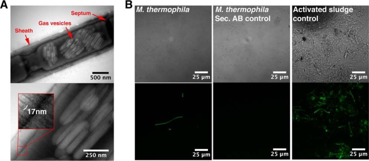
The sheaths on intact filaments display regular striations perpendicular to the filament length and bind the amyloid-specific conformational antibody WO1. A, transmission electron micrographs of intact filaments. The width of the striations is highlighted in the magnified inset. B, binding of the amyloid specific antibody (WO1) to the intact filaments. Differential interference contrast images are shown in the top row, and the green fluorescence signals from the antibodies are shown in the bottom row. Activated sludge from a wastewater treatment plant known to contain both amyloid-positive and -negative cells (40) was used to adjust the settings of the microscope. Secondary controls with only the secondary antibody were included to rule out unspecific binding of the secondary antibody.