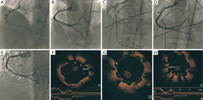
Figure 1.
Diffuse dissection of the proximal to distal RCA (A); stenting of the RCA with six DES (B); very late stent thrombosis of the RCA (C) treated with balloon angioplasty 40 months later (D); patent RCA stents, but diffuse peri-stent staining (arrow) 32 months later (E); OCT showing large arcs of malapposition (arrow) (F); areas of evagination (*) reaching 2.5 mm2 (G); and multiple areas of uncovered stent struts (arrow) (H). RCA, right coronary artery; DES, drug-eluting stents; OCT, optical coherence tomography.

