Figure 5.
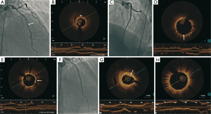
A total of 50% lesion in the proximal LAD and 70% tubular lesion in the mid LAD (arrow) (A); LAD dissection with IMH (*) showed on OCT (B); results after PCI with two DES (C); OCT 15 days later showed patent stents but mild malapposition at the proximal edge of the proximal stent (250 µm) (arrow) (D) and persistent IMH covered by the stents (arrow) (E); 2 years later, 50% in-stent restenosis in the distal segment of the stent (F) with a minimal lumen area of 2.6 mm2 (G) and complete endothelialization of the stent struts (H). LAD, left anterior descending; IMH, intramural hematoma; OCT, optical coherence tomography; PCI, percutaneous coronary intervention; DES, drug-eluting stents.

