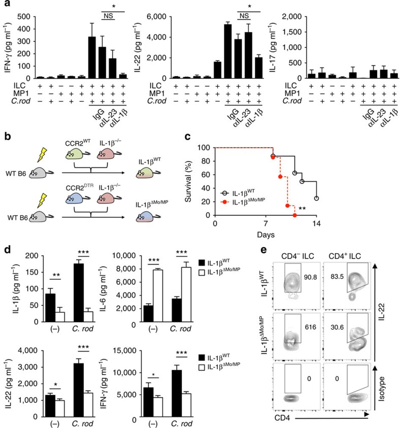
Figure 4. Activation of RORγt+ ILCs requires IL-1β produced by monocyte-derived intestinal macrophages during C. rodentium (C. rod) infection.
(a) CD3-RORγt+ ILCs from uninfected RORγtGFP/+ reporter mice and MP1 cells from C. rodentium-infected CD115GFP were isolated. ILCs and MP1 cells (1 × 10⁶ cells per ml) were cultured alone or co-cultured with or without stimulation with heat-killed C. rodentium (MOI=10) for 24 h. Neutralizing antibodies for IL-23 (10 μg ml−1), IL-1β (10 μg ml−1), or isotype controls were used to block cytokines. Data are given as mean±s.d. (n=4). *P<0.05; NS, not significant by Dunnett's test (compared with isotype control). (b) Schematic illustrating experimental protocol of mixed BM chimera for IL-1β monocyte/macrophage-conditional depletion. Lethally irradiated C57BL/6 recipient mice were reconstituted with mixed bone-marrows from Ccr2WT or Ccr2DTR and Il1b−/− mice (1:1 ratio) for 8 weeks. After 8 weeks, mice were infected with C. rodentium, and CCR2+ monocytes and monocyte-derived MP1 cells were depleted by DT injection (10 ng g−1 body weight) on days 5, 7, 9 and 11 post infection. (c) Survival of chimeric mice infected with C. rodentium (n=7). **P<0.01 by Log-rank test. Results are pooled data of two independent experiments with 3–4 mice each. (d) CCR2WT/Il1b−/− control chimera (IL-1βWT) and CCR2DTR/Il1b−/− chimera (IL-1βΔMo/MP) were infected with C. rodentium and DT was injected (10 ng g−1 body weight) on days 5 and 7. LPMCs were isolated on day 8 post infection. 2 × 10⁶ cells per ml LPMCs were cultured with or without stimulation with heat-killed C. rodentium (MOI=10) for 24 h. Cytokines in the culture supernatant were analysed by ELISA. Data are given as mean±s.d. (n=4, representative of two independent experiments). *P<0.05; **P<0.01; ***P<0.001 by Student's t-test. (e) Isolated LPMCs in d were cultured in the presence of heat-killed C. rodentium (MOI=10) for 16 h. IL-22 production in CD4− ILCs (Lin-Thy-1+CD3−CD4−) and CD4+ ILCs (Lin-Thy-1+CD3-CD4+) was assessed by flow cytometry. Data are representative of four individual mice.

