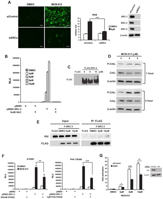
Figure 4. Oxidative stress induced by MCB-613 depends on SRCs and drives further SRC hyper-activation via the Abl kinase signaling pathway.
(A) ROS levels as indicated by CM-H2DCFDA in HeLa cells which were transfected with control siRNAs or siRNAs targeting all three SRCs and treated with MCB-613. A representative picture from multiple fields is shown for each treatment (left panel). Fluorescence signals are quantified (middle panel) and knock down efficiency is shown by immunoblotting (right panel). Scale bar: 200μm.
(B) Luciferase assays on HeLa cells co-transfected with pG5-luc and pBIND or pBIND-SRC-3 expression vectors and treated with increasing concentrations of MCB-613 for 24 hours in the presence or absence of NAC.
(C) The immunoprecipitate from FLAG or FLAG-SRC-3 overexpressing HeLa cells treated with MCB-613 at the indicated concentrations for 1 hour was resolved on a 5% SDS-PAGE gel containing 20 μM Phos-tag and immunoblotted with anti-FLAG antibody.
(D) Immunoblotting of phosphorylated CrkL from HeLa cells treated with MCB-613 for 1 hour or 4 hours.
(E) Immunoblotting of Abl in the coIP complex from FLAG or FLAG-SRC-3 overexpressing HeLa cells treated with MCB-613 at the indicated concentrations for 1 hour.
(F) Luciferase assays on HeLa cells co-transfected with pG5-luc and pBIND or pBIND-SRC-3 and treated with MCB-613 for 24 hours in the presence or absence of AT9283 or PHA739358.
(G) HeLa cells transfected with control siRNA or siAbl1 were transfected with pBIND or pBIND-SRC-3 and pG5-luc, followed by treatment with increasing concentrations of MCB-613 for 24 hours and luciferase assays. Knockdown efficiency of Abl is shown in the right panel.
Data are presented as mean ± SEM. * p<0.05, ** p<0.01, *** p<0.001. See also Figure S4.

