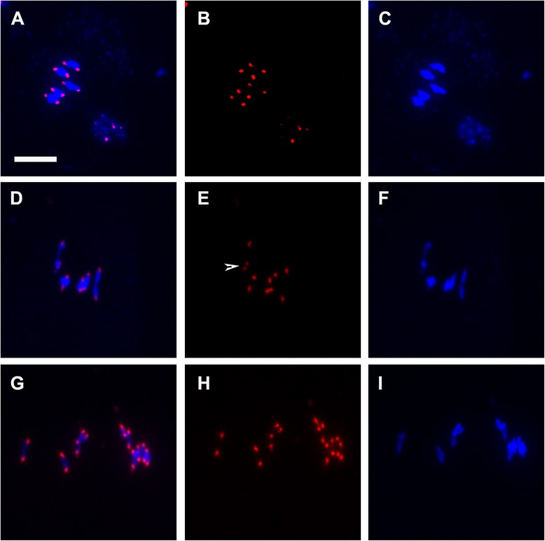
Fig. 2.

Loss of centromeric cohesion in pans1 male meiocytes during Meiosis I. FISH on male meiotic chromosome spreads using a centromeric repeat probe showing DAPI (blue) and probe (red). Left column: merged images of DAPI and the probe; middle column: probe alone; right column: DAPI. a-c wild type metaphase I. d-i pans1 metaphase I. d-f mild phenotype showing split centromere signal on one chromosome indicated by arrow head. (g-i) strong phenotype showing univalents of one chromosome and split centromere signals on four chromosomes. Scale bar 10 μm
