Figure 6.
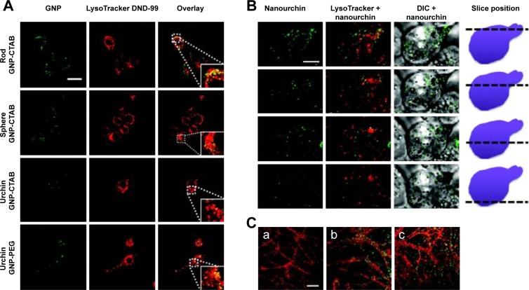
Two-photon luminescence imaging of internalized AuNPs in microglia and primary hippocampal neurons.
Notes: (A and B) Internalization of PEG- and CTAB-coated AuNPs in microglia. AuNPs are primarily found in the cytosol, and to a certain extent, colocalize with lysosomal compartments (indicated by enlarged inset image). Scale bar (20 µm) in control overlay image is representative for all panels. (B) Internalization of urchin AuNPs. Last column of panels provides a schematic illustrating the position along the z axis relative to the cell. Scale bar (5 µm) in the first panel is representative for all panels. (C) Two-photon luminescence imaging of primary hippocampal neurons treated after 21 days in vitro with (b) PEG-coated and (c) CTAB-coated GNPs for up to 4 hours. Neurons were stained with MitoTracker green FM (pseudocolored red). Untreated control cells can be seen in (a). Reprinted with permission from Hutter E, Boridy S, Labrecque S, et al. Microglial response to gold nanoparticles. ACS Nano. 2010;4(5):2595–2606, copyright 2010 American Chemical Society.138
Abbreviations: AuNPs, gold nanoparticles; PEG, polyethylene glycol; CTAB, cetyl trimethylammonium bromide; GNPs, glucose-coated AuNPs.

