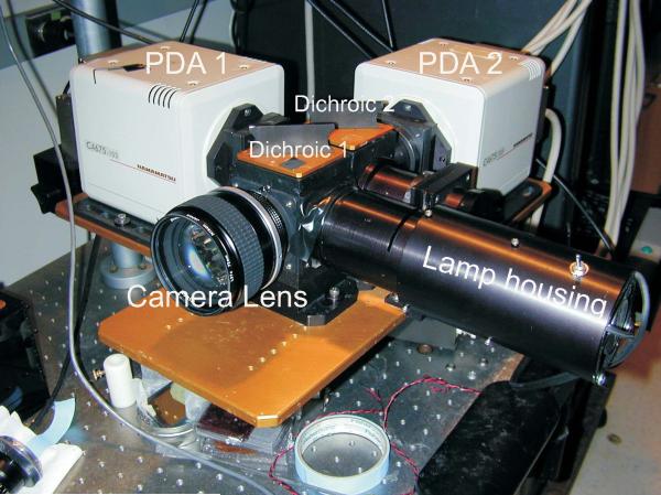
Figure 12.17.1.
A dual optical mapping instrument based on two Hamamatsu photodiode arrays is designed with sealed optics to reduce noise from thermal convection currents in front of the arrays. The photodiode arrays (PDA 1 and PDA 2) and the first-stage amplifiers are housed in the beige boxes where PDA 1 serves to map Cai signals and PDA 2 maps Vm signals. The first dichroic box is located immediately behind the camera lens and splits the excitation beam coming from the lamp housing. The second dichroic box is located behind the first dichroic box and plots the fluorescence from the heart with the low-wavelength image focused on PDA 1 and the long-wavelength image focused on PDA 2.

