Fig. 1.

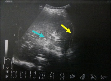
Color doppler ultrasound investigation. The ultrasound showing fluid around liver and hilus lienis (yellow arrow) and uneven right liver lobe (blue arrow). The gallbladder cannot be detected

Color doppler ultrasound investigation. The ultrasound showing fluid around liver and hilus lienis (yellow arrow) and uneven right liver lobe (blue arrow). The gallbladder cannot be detected