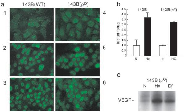
Fig. 2. Functional activity of HIF-1α in ρo cells.

a, nuclear translocation. Wild type (WT) and ρo cells were maintained in normoxia (1 and 4) or exposed to hypoxia (2 and 5) or desferrioxamine (3 and 6) for 2–4 h and immunostained with an anti HIF-1α monoclonal antibody. b, transcriptional activity. Wild type and ρo cells were transfected with a GAL-4-HIF-1α fusion (amino acids 740–826) and a GAL-4-DNA-binding domain luciferase expression plasmid and maintained in normoxia (N) or exposed to hypoxia (Hx). Results are expressed as the mean of arbitrary luciferase units per µg of protein ± S.D. c, RNase protection assay for Northern blot VEGF expression in ρo cells exposed to hypoxia (Hx) or desferrioxamine (Df).
