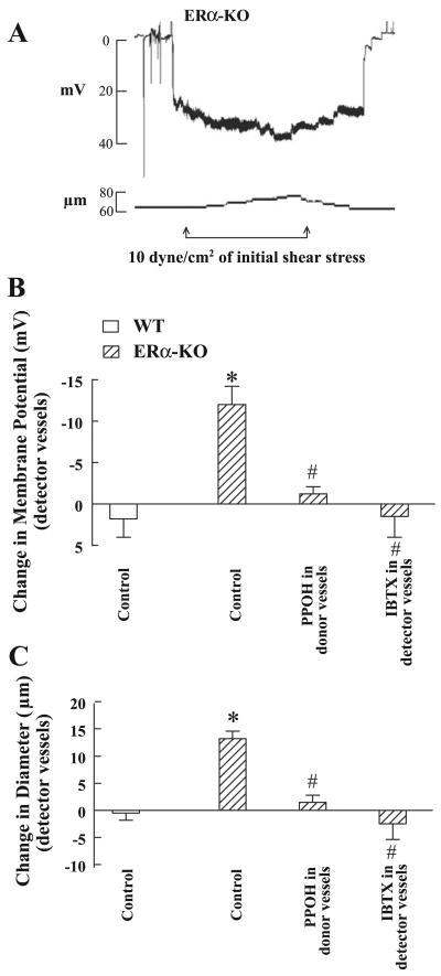
Fig. 2.
A: original tracing of changes in smooth muscle membrane potential (mV) and diameter (μm) of a detector vessel (mesenteric artery) in response to the perfusate from a donor vessel (gracilis arteries) isolated from male ERα-KO mice, stimulated by 10 dyne/cm2 shear stress. B and C: summarized data of changes in smooth muscle membrane potential (B) and diameter (C) of detector vessels of ERα-KO (n = 5) and WT mice (n = 5) in the presence and absence of IBTX or PPOH. *Significant difference from WT control; #Significant difference from ERα-KO control.

