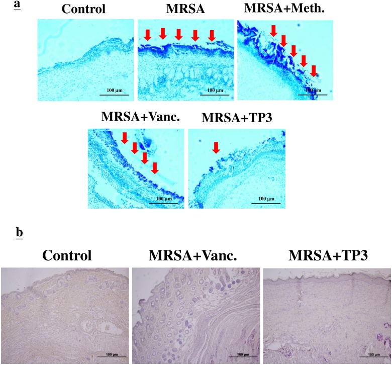
Figure 4. Evaluation of wounds and skin maturation by Gram staining of tissues.
A. Wound biopsy specimens of infected mice (untreated controls or mice treated with the indicated antibiotic or TP3) were Gram stained on day 3. Gram-positive microorganisms are indicated by violet rods. Gram-positive microorganisms were reduced in mice treated with TP3 compared to the untreated group. Arrows indicate Gram-positive microorganisms. The images are representative of two experiments, each performed in triplicate. B. Evaluation of dermal and epidermal maturation. Magnification, x100. The length and height of the photomicrographs are 100 μm.

