
Figure 2. Electron microscopic features of diabetes related clear cell tubules (CCT).
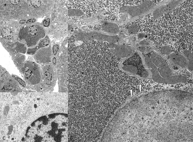
The overview micrograph A. illustrates a transverse section of an altered CCT with enlarged epithelium due to densely packed α-particles of glycogen in the cytoplasm B. in comparison to unaltered distal tubule epithelium C. with small amounts of glycogen and preserved loose distributed cell organelles. In CCT, masses of glycogen displace mitochondria (arrows) to the outer cell borders and the endoplasmatic reticulum (arrowheads) has nearly vanished and is only detectable beneath the nucleus. Length of the lower edge A 50 μm, B 7.5 μm, C 3 μm.