
Figure 3. Autophagic activity in rat clear cell tubulus lesions.
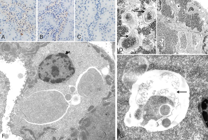
Immunohistochemical demonstration of microtubule associated light chain 3 A (LC3A) indicates autophagy activity in glycogenotic tubules of diabetic rats, without A. or after treatment with NVP/BEZ235 for 4 weeks B. Low autophagy activity was detected in control kidney samples of non diabetic rats following NVP/BEZ235 administration for 4 weeks C. Electron microscopic examination reveals segregation vacuoles in the cytoplasm of the distal tubular epithelium with membrane bound vacuoles (D. arrows), containing glycogen particles, thus representing autophagic vacuoles. In untreated diabetic rats E. they are small and often singular (segmentated square). After treatment with NVP/BEZ235 for 4 weeks F., G. vacuoles are multiple, large, filled with abundant amounts of glycogen and are membrane bound (arrow). Length of the lower edge: A-C 0.15 mm; D 1.5 μm, E 20 μm, F 17 μm, G 7 μm.