
Figure 1. Three checkpoint kinase inhibitors cooperate with gemcitabine to enhance cytotoxicity.
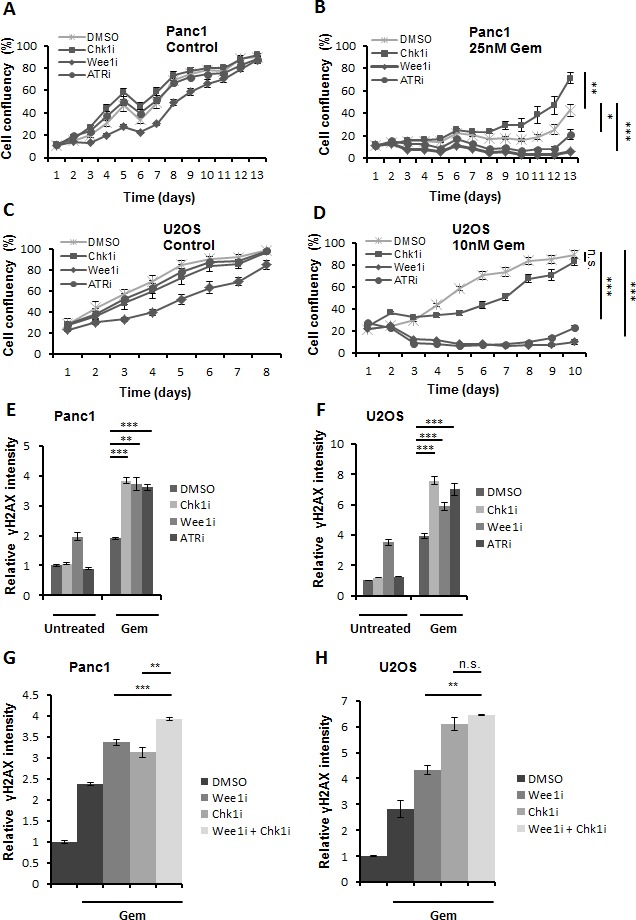
A.-D. Panc1 and U2OS cells were treated with 2.5μM SB 218078, 0.5μM MK-1775 and 5μM VE-821 (referred to as Chk1i, Wee1i, and ATRi, respectively, for their target kinases), in the absence or presence of gemcitabine (Gem) at the indicated concentrations. After 24 h, all drugs were removed and fresh medium was added. Cells were incubated for 8-13 days, and confluency was measured each day using brightfield microscopy (Celigo cell cytometer). Error bars represent the SD, n = 3. p-values (based on Student's t-test, 2-sided, assuming different variances) were determined for the last measurement of respective cell line. E, F. Cells were treated for 24 h with gemcitabine, followed by treatment with checkpoint kinase inhibitors (5μM Chk1i; 1μM (Panc1) or 0.5μM (U2OS) Wee1i; 10μM ATRi) and gemcitabine for another 20 h. Cells were then fixed and stained for γH2AX. Detection and analysis was performed using automated immunofluorescence microscopy (BD Pathway). Error bars represent the SD, n = 3. Images of γH2AX stainings are shown in (Supplemental Figure S4 A, B). G, H. Cells were treated with 1μM Wee1i, 5μM Chk1i or DMSO in the presence of 300nM gemcitabine for 24 h. As a control, cells were treated with DMSO without gemcitabine. The cells were then processed as described in (E-F).