
Figure 2. Inhibition of Wee1 decreases the phosphorylation of Chk1 in gemcitabine-treated cells.
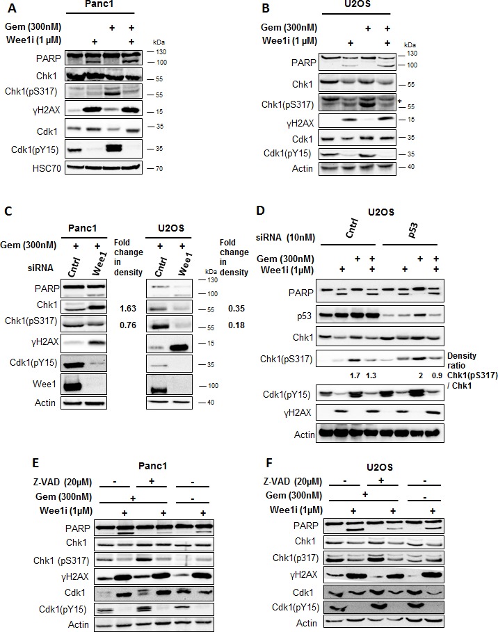
A, B. Panc1 and U2OS cells were treated with 1μM Wee1i or DMSO, with or without 300nM gemcitabine, for 24 h. Blots of cell lysates were stained for phosphorylation of the ATR-substrate Chk1. HSC 70 or β-Actin was stained as loading controls. C. Cells were depleted of Wee1 by transfection with 10nM siRNA for 48h, followed by gemcitabine treatment for 24 h and immunoblot analysis as in (A, B). Scrambled siRNA was used as control. D. Cells were transfected with siRNA against p53 and scrambled siRNA was used as control. After 48 h (for each condition), cells were exposed to Wee1 inhibitor in the presence or absence of gemcitabine. 24 h later, cells were harvested and immunoblotting was performed. β-Actin was stained as loading control. E, F. Cells were treated with Wee1i or DMSO, with or without gemcitabine, in the presence or absence of the pan-caspase inhibitor Z-VAD.fmk at the indicated concentrations. After 24 h, the cells were subjected to immunoblot analysis.