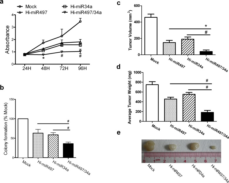
Figure 5. miR-497 and miR-34a synergistically retard cell growth.
(a) The cell-growth curves for A549 cells transfected with Hi-miR497, Hi-miR-34a, or Hi-miR497/34a at 24, 48, 72, and 96 h. Means ± SD, n = 3 (*P < 0.05, at 48 h, Hi-miR497/34a vs. Hi-miR497;#P < 0.01, at 72 and 96 h, Hi-miR497/34a vs. Hi-miR497 or Hi-miR34a). (b) A549 cells were transfected with Hi-miR497, Hi-miR34a, or Hi-miR497/34a. Colony formation was examined in soft agar. Numbers of colonies per well (≥50 cells per colony) in triplicate wells are shown. Mean ± SD (#P < 0.01, Hi-miR497/34a vs. Hi-miR497, Hi-miR497/34a vs. Hi-miR34a). The cooperative effects of miR-497 and miR-34a on tumor formation were examined in a nude mouse xenograft model. Hi-miR497/34a-transfected A549 cells were injected s.c. into the right inguino-abdominal flanks of nude mice. The Hi-miR497/34a-transfected cell treatment generated tumors with smaller volumes (c) and lower tumor weights (d), as determined at necropsy, than those of tumors generated with mock-transfected cells. Mean ± SD, n = 5 (#P < 0.01, Hi-miR497/34a vs. Hi-miR497, Hi-miR497/34a vs. Hi-miR34a). (e) Images show the features of tumor growth at necropsy.

