
Figure 3. MiR-744 functions as a tumor promoter in NPC cells metastasis and growth in vivo.
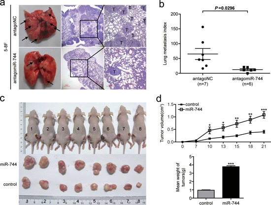
a. Representative photos of mouse lungs and microscopic images of H&E staining of mouse lung sections at 30 days after tail vein injection of 5–8F cells transfected with antagomiR-744 or antagoNC. The black arrows indicate the location of metastatic nodules. T indicates metastatic tumor areas. b. The average of lung metastasis index in the lung from each group of mice. Lung metastasis index was calculated as follows: metastatic tumor areas/total lung areas. c. Subcutaneous tumor growth in mouse xenografts at 21 days after injection with 5–8F cells stably overexpressing miR-744 or control 5–8F cells with stably integrated empty vector. d. Time-dependent tumor growth curves and tumor weights of the different treatment groups. Tumor volume was measured at different time points and calculated with the following formula: V = (L × W2)/2, V, volume (mm3); L, biggest diameter (mm); W, smallest diameter (mm). *p < 0.05; **p < 0.01; ***p < 0.001 compared to controls.