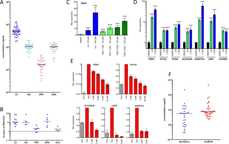
Figure 6. PPARβ/δ ligands are present in ascites at high concentrations and induce PPARβ/δ target genes.
A. LC-MS/MS analysis of polyunsaturated fatty acids (PUFAs) in ascites from ovarian carcinoma patients (n = 38). B. Induction of PDK4 in MDMs after 24 h exposure to different PUFAs in different donors (n = 4-8). Each data point represents a biological replicate. C. Rapid induction (3 h stimulus) of PDK4 by LA and conjugated 9(Z),11(E)-LA and 10(Z),12(E)-LA in MDMs (triplicates). D. Induction of PPARβ/δ target genes in MDMs after 24 h exposure to linoleic acid (LA) in comparison to L165,041 (triplicates). E. Repression of PPARβ/δ target genes in MDMs (n = 3) cultured in ascites for 48 h by different concentrations of PT-S264 added during for the last 24 h of the experiment. Values were normalized to 1 for cells in ascites. F. LC-MS analysis of 15-HETE and the stable prostacyclin derivative 6k-PGF1α in the same samples as in A. Horizontal bars show the medians in panels A and B. Values represent averages of triplicate measurements ± standard deviation in all panels. Significance was tested relative to control cells.

