
Figure 4. Myeloid suppressor cell expansion lasts longer in aged than in young mice after CpG-ODN+IFA treatment.
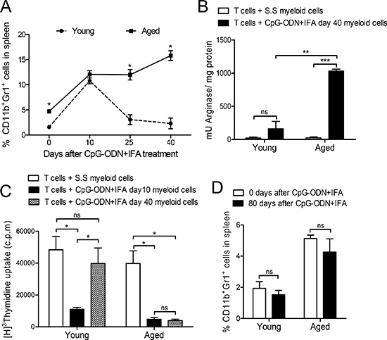
(A) Splenocytes from young and aged mice were obtained 0, 10, 25 and 40 days after treatment, stained with anti-CD11b and anti-Gr1 and analyzed by flow cytometry. (B) Myeloid cells were isolated from young and aged mice 40 days after S.S or CpG-ODN+IFA- treatment and cultured with naïve T cells from young mice stimulated with anti-CD3 and anti-CD28. Cells were prepared for arginase activity analysis. Results are expressed as mU of enzyme activity per mg of protein lysate. (C) Ten and 40 days after treatment, myeloid cells were isolated from spleen of young and aged treated mice and cultured with naïve T-cells from young mice stimulated with anti-CD3 and anti-CD28. T-cell proliferation was measured by [3H]-Thymidine incorporation after 18 h pulse, (values are represented as c.p.m of stimulated minus unstimulated). (D) Zero or 80 days after treatment, splenocytes were removed from mice, stained with anti-CD11b and anti-Gr1 and analyzed by flow cytometry. (A–D) Results are representative of three experiments performed (mean ± SEM; n = 4 mice/group). *p < 0.05; **p < 0.01; ***p < 0.001.