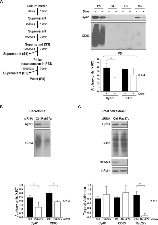
Figure 4. Analyses of cellular and secretome Cyr61distribution.

A. Fractionation of secretome by differential centrifugation from MDA-MB-231-Tet-On-shRNA-c-Src cultures grown in presence or absence of Doxy (2 μg/ml) for 72 h. After protein concentration by methanol/chloroform precipitation of fractions, expression of Cyr61 and CD63 was analyzed by immunoblotting. ImageJ densitometry quantification of four independent experiments (mean ± SD) is reported below and expressed in arbitrary units (**p < 0.01). B., C. MDA-MB-231 cells were transiently transfected with either scramble siRNA (Ctrl) or Rab27a-SiRNA (Rab27a) for 96 h. In secretome fraction S3, obtained from cultures with equal number of cells, and expression of Cyr61 and CD63 determined by immunoblotting B. Expression of Cyr61, CD63 and Rab27a was determined by immunoblotting in total cell extracts. Membranes were reblotted with anti-β-actin for loading control C. ImageJ densitometry quantification of three independent experiments (mean ± SD) is reported below and expressed as Target/β-actin ratio (for total cell extract) or in arbitrary units (for secretome) (*p < 0.05, ***p < 0.001).
