
Figure 5. Exosomes produced by multiple myeloma cells induce osteoclast formation in RAW 264.7 cells and promote formation of lacunae on dentine slices.
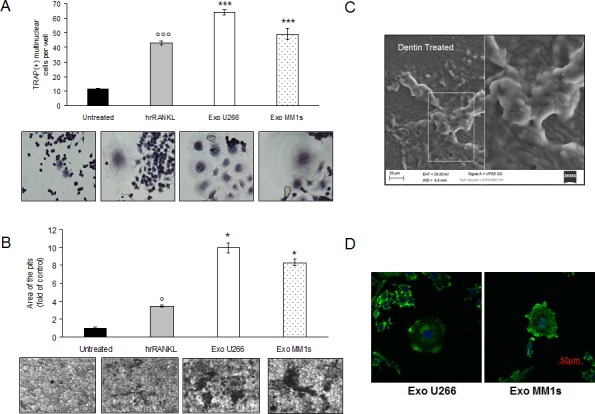
A. Raw264.7 cells incubated with 25ng\ml of hrRANKL (positive control) or with 25 μg/ml of U266 and MM1s exosomes for six days, and then stained for TRAP expression. TRAP positive cells were photographed and counted. Lower panel shows representative pictures of TRAP+Raw264.7 cells (original magnification 20x). Data presented are the mean of three different experiments. Sidak test: *,EXO-U266/MM1s vs Untreated (*p < 0.05); °,hrRANKL vs Untreated (°p < 0.05); B. Bone resorption ability of Raw264.7 cells treated for six days with 25ng\ml of hrRANKL (positive control) or with 25 μg/ml of U266 and MM1s exosomes was evaluated by resorption pit assay on dentine discs. Data relative to the resorbed areas by mature osteoclasts are represented as fold increase of Raw264.7 cells untreated (black column, 1 arbitrary unit). Data presented are the mean of three separate experiments. Sidak test: *,EXO-U266/MM1s vs Untreated (*p < 0.05); °,hrRANKL vs Untreated (°p < 0.05); Lower panel shows representative pictures of lacunae (dark areas) formed by cells treated with hrRANKL or MM-exosomes (original magnification 20x). C. Representative scanning electron microscopy (SEM) image of resorption pits generated by Raw264.7 cells cultured on dentine slices and treated with MM exosomes. Scale bar = 20 μm. D. Confocal microscopy of Raw264.7 cells cultured for six days with 25 μg/ml of U266- or MM1s-derived exosomes and fixed; actin filaments in podosomes were labeled using Alexa Fluor® 488 phalloidin (green) and nuclei were counterstained with DAPI (blue). Scale bar = 50 μm.