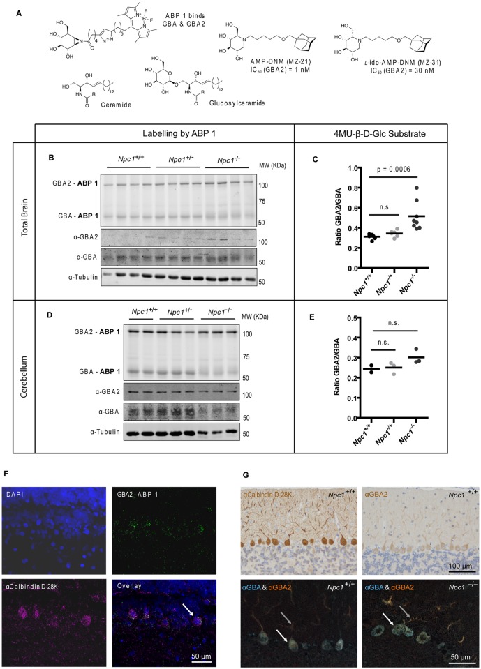
Fig 1. GBA and GBA2 in brain of Npc1 +/+ and Npc1 -/- mice.
(A) Chemical structures of ceramide (Cer), glucosylceramide (GlcCer), the ABP 1 specific for GBA and GBA2, and the inhibitors AMP-DNM (MZ-21) and l-ido-AMP-DNM (MZ-31) with respective IC50 values. (B) Fluorescent labelling of GBA and GBA2 with ABP 1 and Western blotting with anti-GBA2, anti-GBA and anti-tubulin antibodies in brain homogenates of 85-day-old Npc1 +/+, Npc1 +/- and Npc1 -/- mice. (C) Ratio of GBA2 and GBA enzymatic activities (assayed with 4MU-β-D-Glc substrate) in brain homogenates of 85-day-old Npc1 +/+, Npc1 +/- and Npc1 -/- mice. (D) Fluorescent labelling of GBA and GBA2 with ABP 1 and Western blotting with anti-GBA2, anti-GBA and anti-tubulin antibodies in homogenates of dissected cerebella of 75-day-old Npc1 +/+, Npc1 +/- and Npc1 -/- mice. (E) Ratio of GBA2 and GBA enzymatic activities (assayed with 4MU-β-D-Glc substrate) in cerebellar homogenates of 75-day-old Npc1 +/+, Npc1 +/- and Npc1 -/- mice. (F) Fluorescent labelling of GBA2 with ABP 1 (top-right) in wild-type rat cerebellum by ICV injection. GBA was pre-blocked with conduritol-β-epoxide. Sections were also labelled with DAPI (top-left) and immunostained with anti-calbindin D-28K antibody (bottom-left). The arrow indicates a double-labelled cell. Scale bar = 50 μm. (G) Single immunostaining with anti-calbindin D-28K and anti-GBA2 antibodies (top) and double immunostaining with anti-GBA and anti-GBA2 antibodies (bottom) of cerebellar sagittal sections of 85-day-old Npc1 +/+ and Npc1 -/- mice. The arrows indicate PC cell body (white) and dendrites (grey). Scale bars = 100 μm (top) and 50 μm (bottom).

