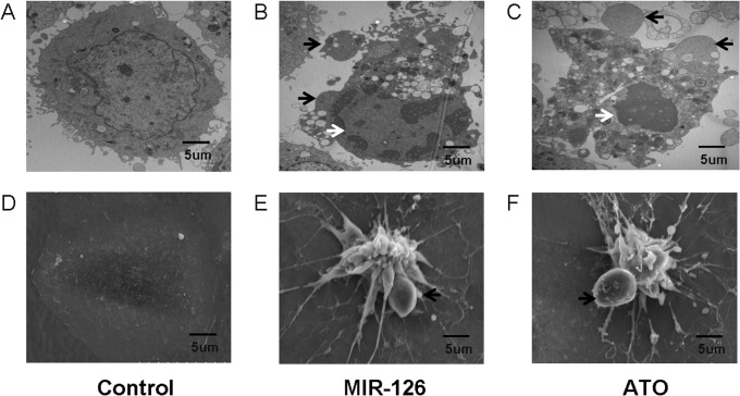
Fig 16. Cell morphology analysis using electron microscopy demonstrates apoptosis.
Induction of apoptosis in HUVECs with post-transfection of miR-126 (B and E), ATO(C and F) and control groups(A and D). Cell morphology alteration including cell shrinkage, generated apoptotic body (black arrows) and nucleus pycnosis (white arrows) were shown by electron microscope assay. TEM (A, B and C) and SEM (D, E and F) images of HUVECs were captured at a magnification of 10,000x.

