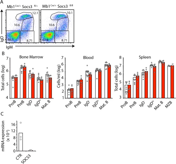
Fig 1. B cell development is independent of SOCS3 signaling.
A, Developing B cell subsets in BM examined by IgM and IgD cell surface expression by flow cytometry. Cells were previously gated as live (DAPI-) CD19+ cells. B, Enumeration of developing B cell subsets in BM (left), blood (center) and spleen (right) of Socs3 +/+ (white) or Socs3 fl/fl (red) Mb1 Cre/+ mice at 8–10 weeks of age. Bars indicate average, circles depict individual mice. Data were pooled from 4 independent experiments. C, Socs3 expression in proB cells from Socs3 +/+ (white) or Socs3 fl/fl (red) Mb1 Cre/+ mice. Expression is relative to Hprt. Bars indicate average, circles depict individual mice. ns (not significant; unpaired two-tailed student’s t test).

