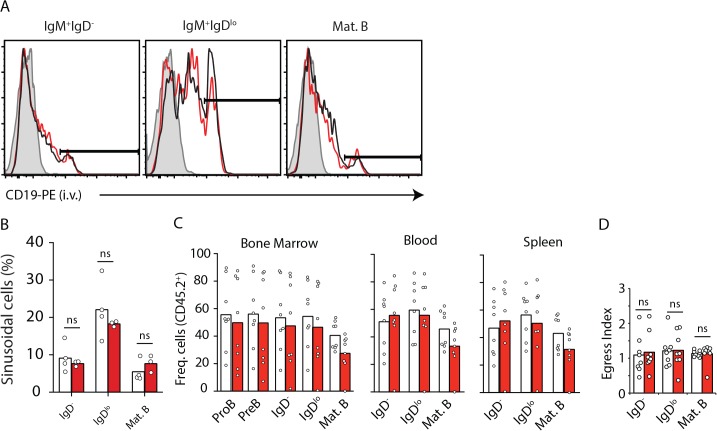
Fig 2. B-lineage cell positioning in parenchyma and sinusoids, and egress from BM is independent of SOCS3 signaling.
A, Distribution of immature IgM+ IgD- and IgM+ IgDlo (both CD93+) cells in BM parenchyma (CD19-PE-) and in sinusoids (CD19-PE+). Histogram of CD19-PE injected i.v. into Socs3-deficient (red) and sufficient (black) mice 2 minutes prior to sacrifice. Filled histogram shows background fluorescence in the indicated B cell subset from a mouse that did not receive CD19-PE. B, Enumeration of immature B cell subsets in BM parenchyma and sinusoids of Socs3-deficient (red) and sufficient (white) mice. Bars indicate average, circles depict individual mice. Data were pooled from 3 independent experiments. C, Frequency of CD45.2+ B-lineage cell subsets in BM (left), blood (middle), and in spleen (right) of lethally irradiated WT mice reconstituted with 50% CD45.2+ Socs3 f/f Mb1 Cre/+ (red) or Socs3 +/+ Mb1 Cre/+ (white) BM cells mixed with 50% CD45.1+ WT BM cells. D, Egress index of B cell subsets from BM calculated as the ratio of CD45.2+ cells in blood by BM. Bars indicate average, circles depict individual mice. Data is representative of three independent experiments. ns (not significant; unpaired two-tailed student’s t test).

