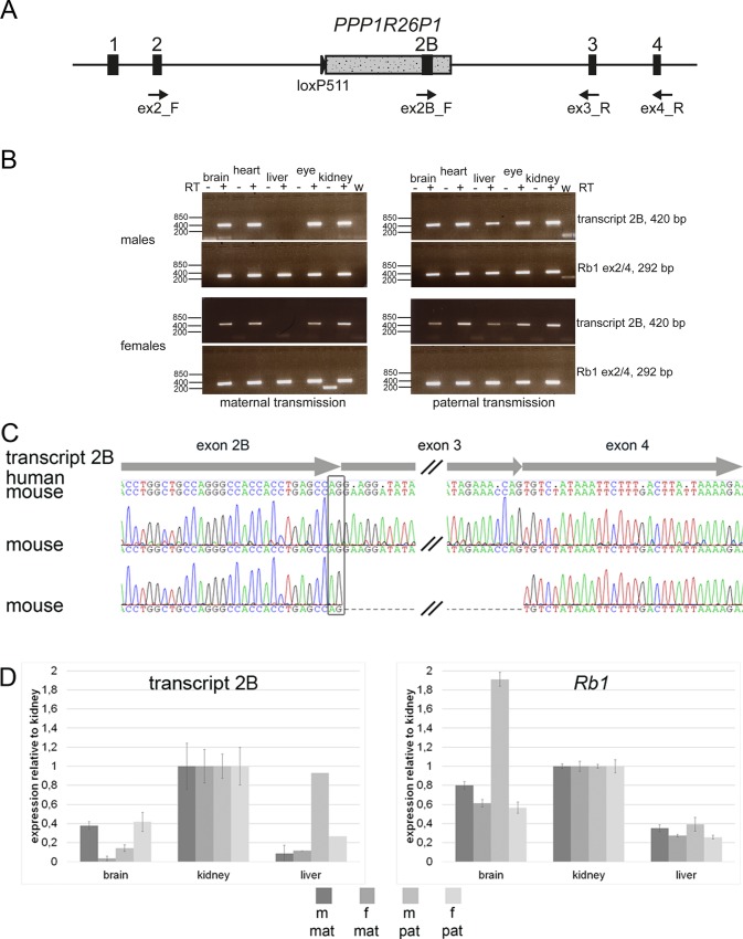
Fig 3. The alternative transcript 2B is expressed in Rb1_PPP1R26P1 knock-in mice.
A) Diagram of Rb1_PPP1R26P1. Positions, names and directions of RT-PCR primers are indicated. B) RT-PCR for transcript 2B (primers ex2B_F / ex3_R, size 420 bp) and Rb1 (primers ex2_F / ex4_R, size 292 bp). +/- RT: with or without reverse transcriptase, w: water control. C) Sanger sequencing of RT-PCR products obtained by reverse priming in Rb1 exon 4 and two rounds of amplification. The alternative exon 2B uses the same splice donor site than in humans, indicated by the black box. Exon structure of transcript 2B is shown with arrows at the top. Consensus sequences of human and mouse transcript 2B are shown with disagreements indicated by dots in the human sequence. Below, two electropherograms are shown of transcripts amplified in Rb1_PPP1R26P1 knock-in mice, the lower showing exon skipping of Rb1 exon 3. D) Quantitative RT-PCR of transcript 2B and Rb1. Mean and standard deviation of four technical replicates are shown. In liver, m/pat and f/pat showed a result for one of the replicates only. m, f: male or female; mat, pat: maternal or paternal transmission of PPP1R26P1.

