
Fig 6. Histologic features of OVA- and sOVA-treated skin sites in BALB/c mice.
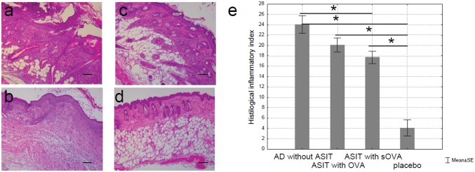
(a) Mice sensitized with OVA and treated with PBS (“AD without ASIT”), (b) mice sensitized with OVA and treated with OVA (“ASIT with OVA”), (c) mice sensitized with OVA and treated with sOVA (“ASIT with sOVA”), and (d) mice sensitized with PBS (“placebo”). Skin sections were stained with H&E and examined at 100x. Scale bars 100 μm. There is marked hyperplasia of the epidermis, a dermal infiltrate (a-c). The cellular infiltrate consists of neutrophils, eosinophils, and lymphocytes. (e) Summary index of the main assessment criteria for histologic skin lesions.