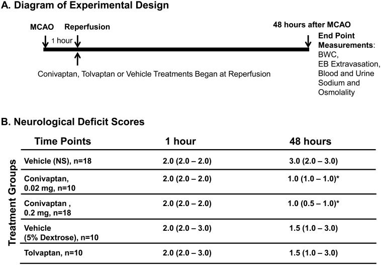
Fig 1. Diagram of experimental design and Neurological Deficits.
(A) MCAO and reperfusion was performed surgically by the intraluminal filament technique. Conivaptan or tolvaptan treatment was initiated immediately at reperfusion (1 hour after MCAO) and lasted for 48 hours. At the end point (48 hours after MCAO) blood and urine was collected for sodium and osmolality measurements, and brains were used to quantify brain water content (BWC) or Evans Blue (EB) extravasation. (B) Neurological deficits were evaluated in all experimental animals twice: before reperfusion (1 hour after MCAO) and at the end point (48 hours after MCAO). Both doses of conivaptan improved NDS at 48 hours after MCAO as compared to vehicle-treated mice. Tolvaptan did not produce any improvements in NDS when compared to the vehicle-treated mice. NDS values are median (25%- 75%), *p < 0.05 vs. vehicle was considered significant.

