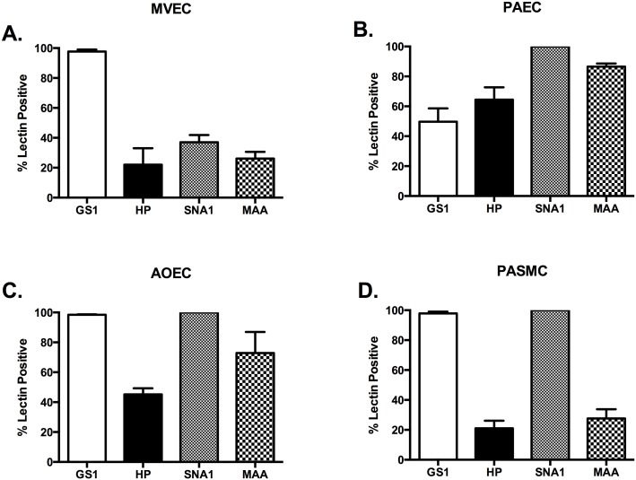
Fig 6. Glycocalyx of cigarette smoke extract injured vascular cells.
All cell types were grown to confluence, media changed to serum free media for 1 hour in the presence of 3% CSE, trypsinized, and stained for our lectin panel in Table 1 as described in methods. (A) MVECs treated with 3% CSE have no significant changes in their glycocalyx profile compared to healthy cells. (B) Injury of PAECs with 3% CSE induces increased lectin binding of GS1 and HP (15.95 ±3.0 vs. 49.67 ±8.92 for GS and 15.58 ± 4.8 vs. 64.47 ± 8.2 for HP), but there was no significant change to SNA or MAA. (C) AOECs treated with 3% CSE had no change in GS1 or SNA1 binding, however both HP and MAA increased significantly (23.48 ± 4.4 vs. 45.20 ± 4.1 for HP and 36.9 ± 1.8 vs. 72.83 ± 14.09 for MAA. P<0.05). (D) There were no significant differences in lectin binding in CSE treated PASMCs.

