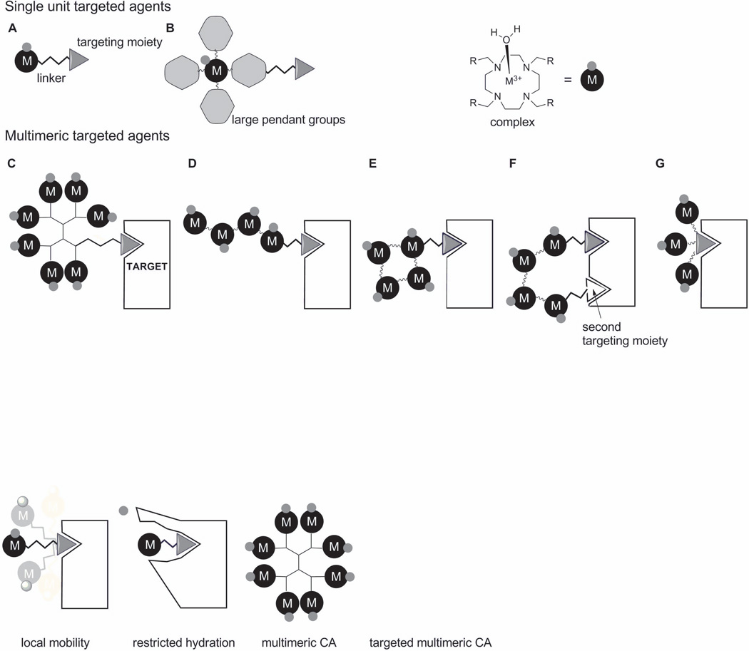
Figure 5.
Designs of monomeric and multimeric contrast agents. A) Simple targeting unit, where the Gd3+ complex is attached to the targeting moiety through a linker. B) The metal ion is placed at the barycenter of a molecule designed to limit molecular rotation. One can also increase sensitivity by increasing the number of Gd3+ ions bound at the target site by use of a dendrimer C) or a straight-chain polymer D). The relaxivity gains due to restriction rotation can be quite small in C) and D) so other approaches to restrict motion by having multiple points of attachment near the target are illustrated in E), F) and G). In these illustrations, the black circled ‘M’s denote ML complexes and the smaller, associated gray circles represent a single water molecule on each chelate.

