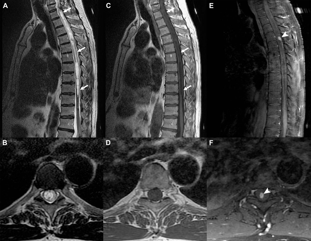
Figure 6.
Gd-based CA for detection of CNS neoplasm. Sagittal and axial T2-weighed (A and B) and T1-weighed (C and D) FSE acquisitions with an extensive thoracic syrinx (white arrows) in a patient with progressive neurological deficits. The etiology remains obscure. Post contrast T1-weighed FSE acquisitions in the sagittal (E) and axial (F) planes demonstrate a small avidly enhancing nodule (arrowheads), which was resected and proven to be a hemangioblastoma (benign hypervascular neoplasm). Without the added information from the CA, the syrinx would be considered idiopathic and no treatment options would be available.

