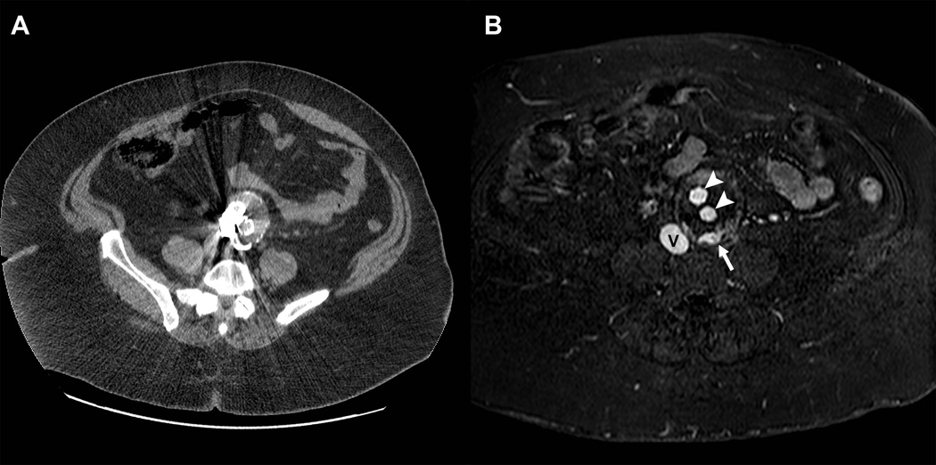
Figure 9.
Clinical application of the blood pool properties of gadofosveset trisodium during follow-up of a patient with an aortic aneurysm. Contrast enhanced computed tomographic scan (A) was of limited diagnostic value due to prominent streak artifacts from metal. GdMS-325 contrast-enhanced magnetic resonance angiography during a blood pool phase acquisition demonstrates excellent signal intensity on the inferior vena cava (V) as well as in the arterial prosthesis (arrowheads).. The white arrow demonstrates an endoleak, which was not detected on CT.

