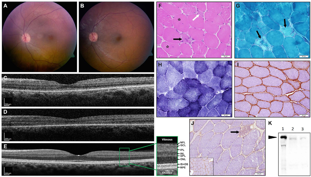
Figure 2. Fundus, OCT imaging, and muscle biopsy results of the affected patients.
(A-E) Fundus imaging: (A) Color fundus picture of the left eye of patient III:3 at age 22 showing a hypermetropic disc with temporal pallor and mild narrowing of the retinal vessels, along with an abnormal macular reflex. (B) Patient III:4 at age 19 shows similar findings with minimal funduscopic changes and preserved retinal structure in the macular area. No pigmentary retinal changes were present in either patient. (C,D) OCT imaging performed at the same time (C- patient III:3, D- patient III:4) revealed retinal structure in the macular area to be largely preserved, with some thinning of the photoreceptor layer (ONL, IS-OS) in the foveal area as compared to an OCT scan of an age-matched normal control (E, retinal layers as seen on OCT are detailed in an enlarged view: NFL- nerve fiber layer; GCL- ganglion cell layer; IPL- inner plexiform layer; INL- inner nuclear layer; OPL- outer plexiform layer; ONL- outer nuclear layer, containing nuclei of photoreceptors; IS+OS- inner +outer segments of the photoreceptors; RPE- retinal pigment epithelium).
(F-K) Muscle biopsy results: (F-J) Staining of muscle biopsy [H&E (F), Modified Gomori-TC (G), NADH (H), and immunohistochemical stains for caveolin-3 (I) and dysferlin (J)]. The results show mild dystrophic changes, including mild variation in myofiber diameter, mild increase of internally displaced nuclei (F, asterisks), and few necrotic (black arrows) as well as regenerating myofibers (F, white arrow). Neither endomysial fibrosis (F) nor significant changes in the cytoarchitecture (H) were observed. There was marked reduction in sarcolemmal staining on immunostain for dysferlin (J, inset-normal control), while immunostain for caveolin-3 (I) was normal. Immunoblot for dysferlin (K, NCL-Hamlet antibody, Novocastra) confirmed marked reduction in dysferlin (arrow indicates dysferlin band (lane 1 - normal control; lanes 2 and 3 - two different loadings of patient’s muscle extract), as opposed to normal immunoblot for calpain-3 (not shown), consistent with a diagnosis of dysferlinopathy.

