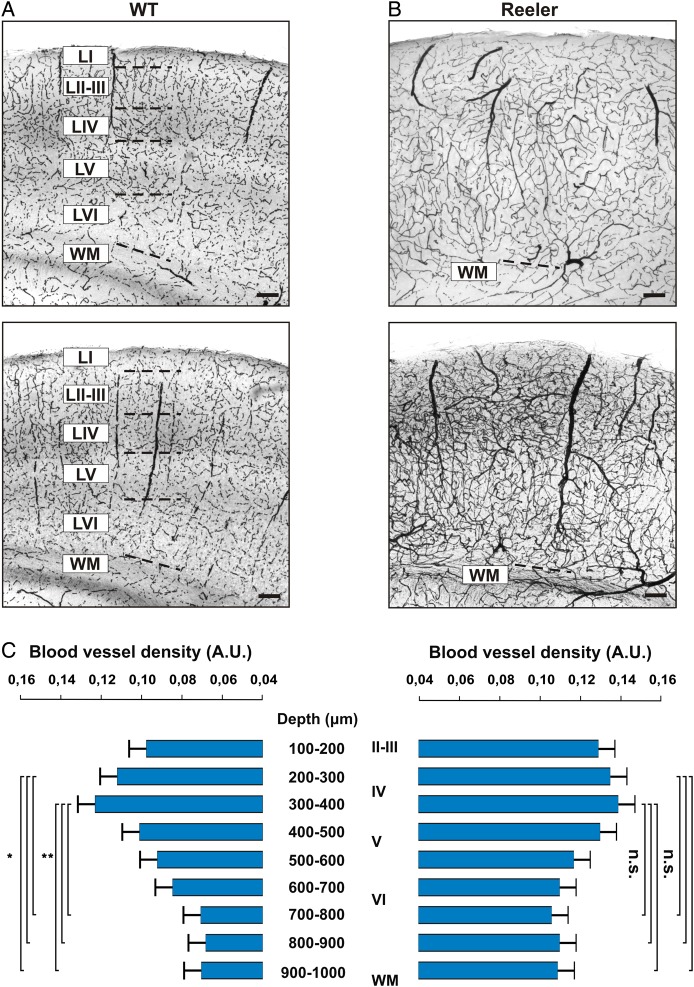
Figure 5.
Vascular network in the barrel cortex of WT and reeler mice. (A and B) Coronal sections through the somatosensory cortex of 2 representative WT and reeler mice, respectively (n = 6 in each group). DAB staining was used to stain erythrocytes in an unperfused brain, providing an indirect but efficient way of revealing the vascular network of the somatosensory cortex. wm: white matter. Scale bar: 100 μm. (C) Relative density of blood vessels as a function of depth in WT (left) and reeler (right) animals. Density was calculated in 100 μm bins from the pia to the white matter. The first 100 μm below the pia were excluded because a heightened background was consistently found near the pia.

