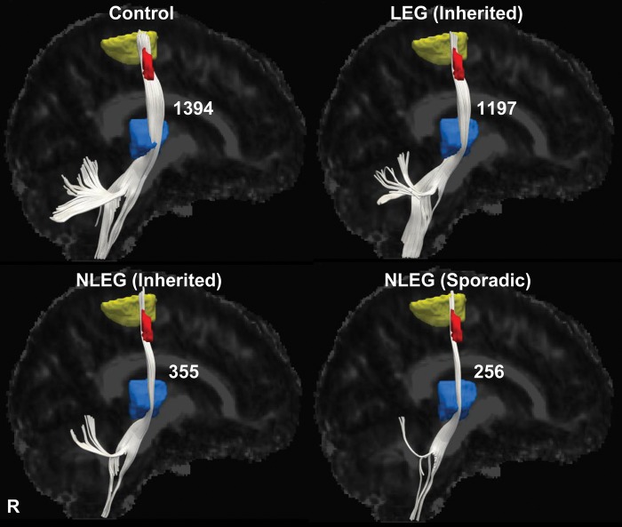
Figure 4.
Thalamocortical tract differences related to phenotype. Display of reconstructed thalamocortical fiber tracts from patients with inherited (LEG and NLEG) and sporadic (NLEG) dystonia and from healthy volunteer subjects. Projection pathways were reconstructed using the subrolandic lower limb-related FA cluster (red) as the seed region and the corresponding motor cortical (yellow) and ventral thalamic (blue) fMRI activation clusters as the target regions (see Materials and Methods). Thalamocortical fiber numbers were preserved in the LEG dystonia subjects (−14%, relative to the healthy control group), but were reduced in both inherited and sporadic NLEG dystonia patients (−75% and −82%, respectively). [The fiber number for each group is presented to the right of the reconstructed fiber tract].

