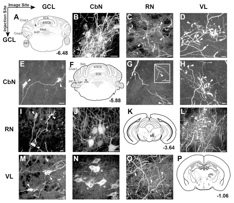
Figure 2.
Consistent structures were labeled along the course of CbN output neurons (columns) following BDA or viral injections into various sites along pathway (rows), including cerebellar cortex around the base of the Simple lobule, CbN, red nucleus (RN), and ventrolateral thalamus (VL). A–D: Row 1 shows somatic BDA label in the CbN (B), boutonal label in the RN (C) and boutonal label in the VL (D) following injections into the cerebellar cortex granule cell layer (sketched in A) (granule cell layer injection just posterior to Simple lobule, at the base between 4/5 and 6 lobule; n=5): E–H: Row 2 shows labeled mossy fiber rosettes in granule cell layer of 4/5 lobule (E); a sketch of the injection site (n=5) (F); labeled boutons in the RN (G) and labeled boutons in VL following injections into the anterior interposed nucleus (H). I–L Row 3 shows GFP expression in mossy fibers (I); GFP expression in somata of the interposed nucleus (J); a sketch of the injection site (K) and boutonal GFP expression in VL (L) following virus injections to the RN of Ntsr1-cre mice (n=5): M–P: Row 4 shows labeled mossy fibers (M); somata in anterior interposed nucleus (N); terminal boutons in the RN (O) anda sketch of the injection site (P) following VL injection (n=11). Scales: B,J,N= 20 μm; C,D,E,G,H,I,L,M,O = 10 μm. Markers: Arrows, axons; Arrowheads, boutons; Asterisks, somata; Crosses, injection sites.

