
Figure 3.
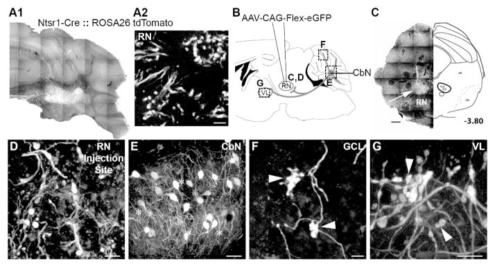
Genetically identified cerebellar nuclear (CbN) neurons branch to innervate red nucleus (RN), ventrolateral thalamus (VL) and 3, 4/5, Simple, Crus I, Crus II, paramedian and copula pyramis lobules of the granule cell layer. A. Transgenic mice (Ntsr1-Cre::Rosa26-tdTomato) show Cre expression in CbN somata (A1) but not RN somata (A2). B. Schematic of AAV1-CAG-Flex-eGFP illustrates injections into the RN of Ntsr1-Cre mice. Tissue was analyzed for terminal label in the granule cell layer, somata label in the CbN and terminal label in the VL (dashed boxes) as well as elsewhere in midbrain and brainstem. C. RN injection site was marked by labeled terminal boutons after viral injections (left). An atlas section identifying RN approximately 3.80 mm posterior to bregma is included for context. Asterisks indicate injection sites for each animal (right). D. Higher magnification image of terminals in the rRN at the injection site confirm viral transfection of Cre-expressing axons, not RNsomata. E. Somata in the interposed nucleus express GFP following viral transfection of RN in Ntsr1-Cre mice. F. Mossy fiber rosettes in the granule cell layer (arrowheads) express GFP. G. Terminal boutons (arrowheads) in VL express GFP following RN viral injections. Scales: C = 500 μm; D, G = 20 μm; E = 50 μm; F = 10 μm.