Figure 8.
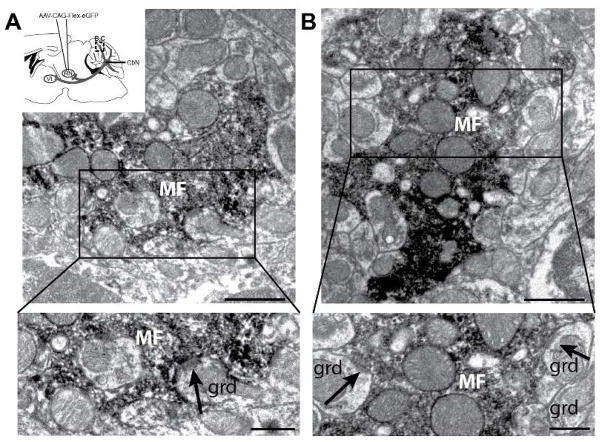
Nucleocortical terminal active zones are adjacent to granule cell postsynaptic densities. A. (inset) Schematic diagram showing AAV1-CAG-Flex-eGFP injection to the red nucleus of Ntsr1-Cre mice. Tissue was analyzed for nucleocortical collateral terminal label in the granule cell layer (dashed box). GFP was labeled with DAB for visualization with electron microscopy. A, B. (top) Electron micrographs showing DAB-labeled mossy fiber rosettes interdigitating with putative granule cell dendrites (grd). Scale = 1μm; (bottom) Mossy fibers in regions boxed above contained vesicles apposed to postsynaptic density on putative granule cell dendrite (arrows). Scale = 0.5μm.

