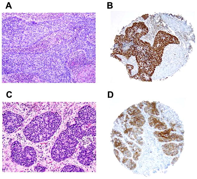
Figure 2. Basaloid pattern in squamous cell carcinoma and large cell neuroendocrine carcinoma (original magnification, x100: A–B).
(A) Basaloid squamous cell carcinoma showing prominent peripheral palisading of tumor cells with scanty cytoplasm. (B) Basaloid squamous cell carcinoma positive for p40. (C) Large cell neuroendocrine carcinoma showing basaloid-like pattern with rosette-like features and nuclear palisading. (D) Large cell neuroendocrine carcinoma positive for CD56.

