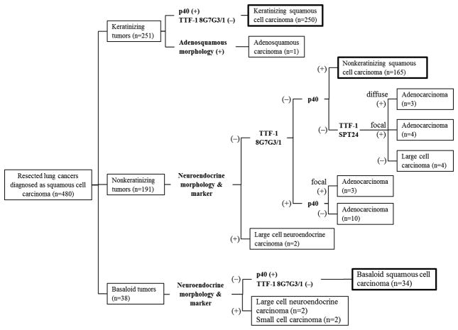
Figure 3. Flowchart reclassifying tumors originally diagnosed as squamous cell carcinoma (n=480) with histologic review and immunohistochemical analysis.
Among keratinizing tumors (n = 251), 250 were confirmed as keratinizing squamous cell carcinoma—p40 positive but TTF-1 (8G7G3/1) negative—and one tumor was reclassified as adenosquamous carcinoma since it had both squamous cell carcinoma and adenocarcinoma morphology. Among nonkeratinizing tumors (n = 191), 165 cases were confirmed as nonkeratinizing squamous cell carcinoma—p40 positive but TTF-1 (8G7G3/1) negative—and there were 20 cases reclassified as adenocarcinoma since they were TTF-1 (8G7G3/1 or SPT24) positive but p40 only very focally positive or negative. The two cases reclassified as large cell neuroendocrine carcinoma exhibited neuroendocrine morphology and differentiation. Among basaloid tumors (n = 38), 34 were confirmed as basaloid squamous cell carcinoma—p40 positive but TTF-1 (8G7G3/1) negative—and 4 tumors were reclassified as high-grade neuroendocrine carcinoma (2 large cell neuroendocrine carcinomas and 2 small cell carcinomas) because they exhibited neuroendocrine morphology and differentiation.

