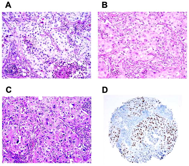
Figure 4. Squamoid morphology of lung adenocarcinoma (original magnification, x100: A–C).
(A) Cytoplasmic keratinization-like feature (eosinophilic cytoplasm with pyknotic nuclei) identified near necrotic area (bottom right). (B) Squamoid tumor cells that exhibited abundant eosinophilic cytoplasm. (C) Squamoid tumor cells the exhibited abundant eosinophilic cytoplasm with sharp cell borders and intercellular bridge-like features. (D) Squamoid adenocarcinoma positive for TTF-1 (8G7G3/1).

